
Women Get Ultrasounds Instead of Mris During Pregnancy Because This Is What a Fetus Looks Like in an MRI
Picture this: you’re scrolling through social media and stumble across a black-and-white scan of a baby in the womb—only it doesn’t look serene or soft like those grainy ultrasound photos we’re used to. Instead, the image is eerie, skeletal, and intense—almost alien. It’s real. And it’s from an MRI.
It’s hard to unsee something like that. For many, it sparks discomfort. For others, curiosity. Why does it look so different? And more importantly, why don’t we see more of it? Every year, millions of expectant parents get their first glimpse of new life through an ultrasound. But if MRI images offer far more detail, why aren’t they the standard? Is it safety? Cost? Something else entirely?
Ultrasound vs. MRI: What’s the Difference?
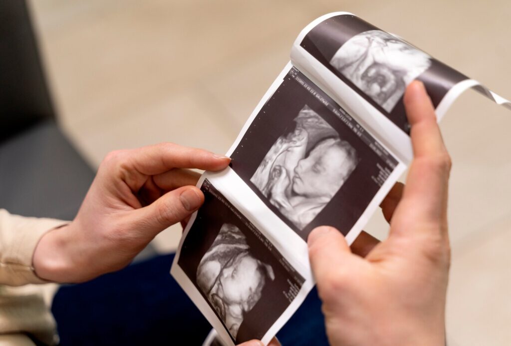
Imagine standing in a dark room with only a flashlight. You can sweep the beam across the space, catching the general shape of things—soft edges, some movement, a sense of life. That’s ultrasound. It sends out high-frequency sound waves, which bounce off structures in the body and return to create a moving image. It’s real-time, safe, and non-invasive, which is why it’s been the go-to tool in prenatal care for over half a century. Ultrasound allows doctors—and parents—to watch a fetus kick, stretch, or even suck its thumb. It’s more than just a medical tool; it’s the emotional entry point into pregnancy for many. The pictures are soft, grainy, and familiar, offering just enough clarity to make out features, while leaving room for imagination and awe.
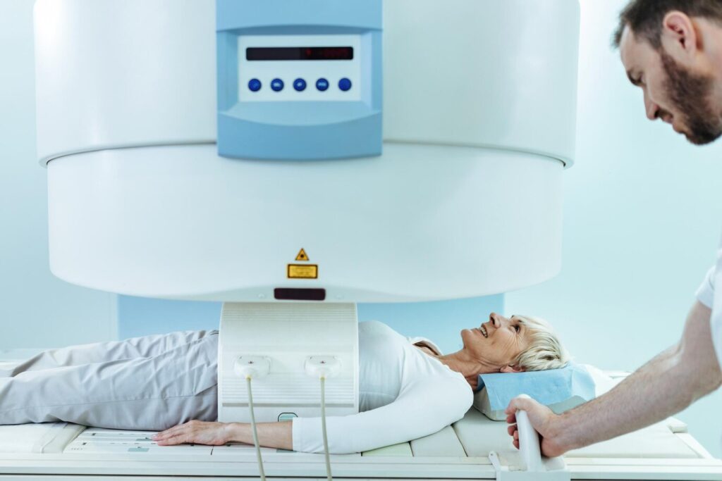
MRI, on the other hand, is a different kind of lens. It doesn’t use sound—it uses powerful magnets and radio waves to capture the body’s internal structures in stunning detail. If ultrasound is a flashlight in the dark, MRI is flipping the room lights on. You see everything. Cross-sections of the brain. The curvature of a forming spine. The chambers of the heart. The level of detail is almost surreal. It can even show subtle abnormalities in tissue development that an ultrasound might miss entirely. For certain conditions, like suspected brain malformations or congenital defects, MRI provides doctors with the clarity they need to make informed decisions and offer critical care.
But this level of clarity comes with a cost—not just financially, but emotionally. The images captured in an MRI scan aren’t softened by movement or blurred edges. They can look harsh, skeletal, even unsettling to someone who isn’t trained to read them. And while that might seem like a superficial concern, it speaks to something deeper: how we emotionally process the idea of life before birth. Science gives us tools, but how we use them—and how we respond to what they show us—is shaped by more than just technology. It’s shaped by feeling, familiarity, and the way we’re wired to make sense of what we see.
Why Ultrasound Is the Go-To for Pregnancy

Ultrasound has earned its place as the default imaging method during pregnancy not just because of what it shows, but because of how seamlessly it fits into the process of care. It’s fast, affordable, and widely available. It doesn’t require specialized environments, and the scans can be done in routine clinical visits, even in low-resource settings. That accessibility has made it an indispensable tool across the globe. More importantly, it provides just enough visual clarity to assess fetal growth, check for abnormalities, and reassure both doctors and parents—all without overwhelming detail. It’s a glimpse, not a dissection. And for most pregnancies, that glimpse is more than enough.
There’s also the fact that ultrasound is incredibly safe. It uses high-frequency sound waves—no radiation, no magnetism, no injections or sedatives. The procedure doesn’t require the fetus to stay still, and it poses no discomfort to the mother. That low barrier to use makes it ideal for routine monitoring, especially during key moments like the first heartbeat, the gender reveal, or the third-trimester growth check.
Over decades, countless studies have affirmed its safety, giving it a track record that builds both clinical trust and emotional comfort. It’s become so standard, in fact, that many parents don’t question it—they simply expect it, and welcome the familiarity it brings.
But maybe the strongest reason ultrasound remains so dominant is emotional. Those grainy, moving images on the screen have become cultural milestones. They’re printed and framed, sent to grandparents, posted online. They make something invisible feel real. They create connection. And while they might not offer the fine-grain anatomical details of an MRI, they offer something else: intimacy. The blurry outlines of a foot, the gentle flutter of a heartbeat—these moments speak to us on a human level. They don’t just confirm a pregnancy; they begin a relationship.
So Why Use MRI at All?
Even though ultrasound covers most of what’s needed in a typical pregnancy, MRI becomes vital when something isn’t adding up. If an ultrasound flags a potential issue—maybe a concern with the brain, spine, or internal organs—doctors may recommend a fetal MRI for further evaluation. That’s when MRI shifts from being a curiosity to a life-saving tool. Its ability to create detailed, high-resolution images of soft tissue allows medical teams to see the fetus with a precision that ultrasound simply can’t match. In complex cases, this added information can mean the difference between early intervention and a missed opportunity. It can guide surgical planning, prepare neonatal teams, or help determine whether a condition is treatable or life-threatening.
Despite some lingering public confusion, MRI is considered safe during pregnancy. It doesn’t expose the fetus to ionizing radiation, and the magnetic fields used in MRI haven’t been shown to cause harm. In fact, many high-risk pregnancy centers use fetal MRI routinely when needed, especially in the second and third trimesters. That said, it’s not used lightly. It’s more expensive, it takes longer, and it requires the pregnant person to remain still inside a loud, narrow tube for an extended period of time—sometimes uncomfortable, always demanding. That’s part of the reason why MRI is reserved for specific situations. It’s not about risk; it’s about necessity.
There’s also a deeper ethical layer to consider. With MRI’s clarity comes emotional weight. Some parents may face painful decisions based on what those scans reveal. Others may carry a diagnosis that has no treatment but still changes how they experience the rest of the pregnancy. So even though the tool is safe, the information it uncovers can carry heavy implications. That’s not something to fear—but it does require compassion, context, and care. The power of MRI lies in its ability to inform, not just impress. It’s a tool of precision, and with that precision comes responsibility—both in how it’s used and how its images are shared with those outside the clinical space.
What Does a Fetal MRI Actually Look Like?
The real reason they discourage MRIs during pregnancy is because then people would realise they’re incubating nightmare demons and would be rightfully terrified pic.twitter.com/55zEeOofsP
— Katie (@ZiziFothSi) May 19, 2021
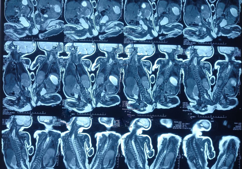
If you’ve only seen fetal ultrasounds, the first glance at an MRI image can be jarring. Instead of soft gray shapes, you’re met with anatomical detail: hollow eye sockets, skeletal outlines, thin slices of the body like pages in a book. These aren’t the dreamy visuals we associate with pregnancy—they’re scientific. Fetal MRI captures cross-sectional views of the body in extremely high resolution, showing layers of tissue, developing organs, and the internal layout of a tiny human. For someone untrained, it can seem harsh, even eerie. But for doctors, this clarity is essential. It allows them to evaluate structures as delicate as brain folds or spinal cords, to look for signs of rare conditions, and to do it all without harming the fetus.
What makes these images so powerful—detail, contrast, accuracy—is also what makes them unsettling for people scrolling past them on social media. We’re not used to seeing developing life through such a clinical lens. And without the warm framing of a doctor or a comforting environment, the images can easily be misinterpreted. A fetal MRI stripped of context becomes an object of shock or curiosity online, labeled as “creepy” or “demonic” by people who don’t understand what they’re seeing. But that shock says more about us than it does about the scan. We’ve become accustomed to romanticized versions of pregnancy—images that are soft, distant, idealized. MRI pulls us out of that space and into something far more raw.
But within that rawness is something remarkable. The MRI isn’t just about exposing the fetus—it’s about honoring the complexity of life. Each scan is a testament to how intricate human development is, how early the brain starts to take shape, how the body assembles itself organ by organ. It’s not a distortion—it’s a deeper reality. And for medical teams, this view can be the difference between a vague suspicion and a clear diagnosis. For all its clinical sharpness, the fetal MRI is deeply human. It’s a window into the beginnings of life—just one we’re not used to peering through.
Why Context and Emotion Matter in Medical Imaging
Images don’t just show us facts—they shape how we feel. And when it comes to pregnancy, emotions run deep. We’re conditioned to expect certain visuals: soft curves, peaceful profiles, the grainy magic of an ultrasound photo taped to the fridge. So when something breaks that pattern—when we see a fetal MRI instead of an ultrasound—it’s not just the image that surprises us. It’s the disruption of what we emotionally associate with that moment. That disruption can lead to discomfort, and that discomfort, if left unexplored, can turn into fear or misunderstanding. This is why context is everything. An MRI image without an explanation can trigger the wrong story in someone’s head.
Medical professionals understand this. Many radiologists and obstetricians take time to walk patients through what they’re seeing and why it looks the way it does. They know that a skeletal-looking face or an empty eye socket isn’t a sign of anything wrong—it’s just how the human body looks under certain imaging conditions. But when these images are shared outside the exam room—especially online—they’re often divorced from that vital context. And in the absence of explanation, the mind fills in the blanks. That’s where myths grow. That’s where fear takes root. What’s intended to diagnose and inform becomes a source of anxiety, simply because it wasn’t introduced with care.
But there’s also hope in this moment. These reactions, while emotional, also represent a deep curiosity—a desire to understand what we’re seeing. And that’s the opportunity: to shift the narrative, to turn fear into education, to help people see not just what’s unfamiliar, but what’s extraordinary. Medical imaging can be cold, yes, but it can also be illuminating. It can show us how fragile and brilliant life is at the same time. If we take the time to listen to what the science is showing us—not just react to how it looks—we open the door to a deeper appreciation of the human experience, in all its forms.
What We See vs. What’s Real

An image is never just a picture. It’s a reflection of how we think, what we value, what we’re afraid of, and what we hope for. When we look at a fetal MRI and recoil, we’re not responding to danger—we’re responding to unfamiliarity. We’re seeing life in a way we haven’t before: exposed, layered, raw. But within that rawness is the truth of who we are. The details we’re not used to—the hollow eyes, the segmented limbs, the fine threads of developing tissue—aren’t errors or horrors. They’re biology. They’re beauty. And when we take a moment to sit with that, to let our minds catch up to what the science is showing us, we start to see something deeper than fear. We start to see awe.
This isn’t about choosing ultrasound over MRI, or tradition over innovation. It’s about understanding that each tool gives us a different lens into life. One is gentle and familiar. The other is precise and profound. Both serve a purpose. Both are essential. And both deserve to be understood for what they are—not just in clinical terms, but in emotional terms, too. The real challenge isn’t in choosing the right tool. It’s in learning how to interpret what we see with empathy, curiosity, and humility.
So the next time you see one of those stark fetal MRI images floating around the internet, take a breath. Step back. Ask not just what am I looking at, but why does it make me feel this way? Because in that question lies the bridge between science and story, between data and meaning. And it’s in that space—where clarity meets compassion—that the real transformation begins.
Featured Image Source: Shutterstock